How do human pathogens adapt? New Wake Forest research holds some clues
HIGHLIGHTS
- Chemistry researcher Rebecca Alexander studies an unusual bacteria that penetrates and lives inside human cells.
- This pathogen keeps a double-sized protein that may help it survive and reproduce.
- New research uncovering this unique bacterial structure could introduce a drug therapy target.
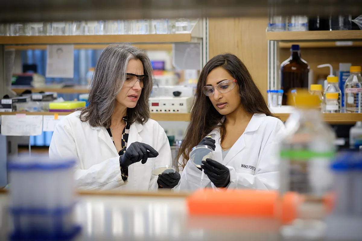
Research underway in the lab of Rebecca Alexander, professor in Wake Forest University’s Department of Chemistry, is shedding light on a peculiar human bacteria, Mycoplasma penetrans.
Alexander’s work to understand how and why this unique pathogen acts as it does could one day lead to new classes of drugs that better target illnesses associated with this particular pathogen, as well as others that act in a similar way.
The research “just expands the possibilities of anti-bacterial therapies,” said Alexander, also a senior associate dean for research and community engagement. “M. penetrans is weird because it actually penetrates into the host cell itself. It’s an intracellular, parasitic pathogen. These bacteria have a sort of spear on the end that they insert into the human host, and then they live inside the host cell. That makes it more like a virus, because that’s how viruses work. Most bacteria don’t do that, but this bacteria does.”
How does this unusual pathogen reproduce?
Her research delves into the intricacies of cell biology—how cells, and specifically M. penetrans, carry out key functions. This unusual pathogen, first discovered as a co-infection in immunocompromised HIV patients, possesses a very small genome, or the collection of DNA it needs to survive. M. penetrans relies on the human host for many genes it doesn’t contain itself.
In the cell, messenger ribonucleic acid (mRNA) carries genetic instructions from the DNA inside the nucleus to other parts of the cell.
Another molecule, transfer RNA (tRNA) reads the instructions on the mRNA molecule and brings specific amino acids together to form peptide bonds. Those bonds build full proteins for cellular structure and function. Peptides do things like regulate blood sugar and reduce inflammation.
There are about 20 standard amino acids and corresponding families of tRNAs. Specific enzymes are required to accurately and rapidly attach the correct amino acid to its tRNA partner. The particular enzyme Alexander has studied for several years is methionyl-tRNA synthetase, or MetRS.
Understanding the cell’s survival strategy
M. penetrans is odd because the MetRS enzyme responsible for attaching the amino acid called methionine to tRNA is twice as large as the equivalent protein found in the common bacteria E. coli. Although bacteria generally shed unnecessary genes, M. penetrans retained this double-sized protein because it appears to perform a separate enzymatic activity that synthesizes methionine.
This might help the pathogen survive when methionine is scarce in the host cell.
In her research extending back more than a decade and in the latest published in PLOS One, Alexander and her co-authors found that this double-sized M. penetrans enzyme has two functions that might work to the benefit of the pathogen.
“The more we can understand about pathogenic microbes, like bacteria or viruses, and how their essential machinery is different, that makes it more drug worthy,” Alexander said, adding that the way M. penetrans functions appears to be unique.
“It almost seems like it was an accident of evolution, but this bacteria said, ‘Oh, that’s actually useful for me, so I’m going to hang onto it.’ So, having the extra domain now gives us a very distinct target for inhibiting this pathogen.”
Wake Forest ties strengthen research
To more fully understand the how and why of M. penetrans MetRS requires an understanding of its structure. A collaboration to tackle that challenge actually involved 1997 Wake Forest alumna Elizabeth Stroupe, now a professor of biological sciences at Florida State University.
Stroupe’s connections to the University extend back to her grandfather, Henry Stroupe, a Wake Forest professor of history and founding dean of the Graduate School of Arts and Sciences. Her collaboration with Alexander brings an expertise in mapping the structure of M. penetrans using a technique called cryo-electron microscopy.
Alexander and Stroupe co-authored the recent PLOS One publication with their graduate students.
“Having the structure of M. penetrans MetRS helps us take the next step of understanding the function better,” Alexander said, adding that she and Stroupe are looking for new methods to more comprehensively model the complex structure of this unique pathogen.
— Contributed by Elaine Ellis
Recent News

Solar energy center begins operations with support from Wake Forest University
Solar energy center begins operations with support from Wake Forest University
Sabin Center conference explores leading in times of environmental crisis
Sabin Center conference explores leading in times of environmental crisis